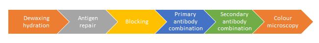
Immunohistochemistry
(IHC) is a technology applying the antigen-antibody
specific binding principle to determine the position, qualitative and
quantitative properties of intracellular antigen (polypeptide and protein)
through the colorimetric chemical reaction of the labelled antibody color
reagent (fluorescein, enzyme, metal ion, isotope).
Immunohistochemistry
is not difficult, but it's not easy to make beautiful dyeing results. The key
to success is that immunohistochemistry detail decides success or failure. what
is the "details"? Here are the details and process. (Paraffin tissue
section as an example)
1.Specimen fixation
The purpose of fixation is not only to solidify protein
in cells, to reduce or terminate the reaction of endogenous or exogenous
intracellular decomposing enzymes, but also to prevent autolysis of tissue
cells, so as to preserve the antigenicity of tissues or cells, so that antigens
do not lose activity and or disperse. The tolerance of different antigens to
the fixed solution is different, so the appropriate fixative should be chosen according
to the antigens.
Now the commonly used fixers are neutral formaldehyde, 4%
polyformaldehyde - phosphate buffer. Some fixed agents have better effect on
special organization, for example, picric acid has a fixed effect for softening
scalp and nails, Helly is better for fixing islet and pituitary, PLP is better
for fixing liquid sugar and preserved tissue antigen.
2. Dehydration, paraffin embedded and cut into slices
Dehydration with gradient ethanol (low to high) and full
dehydration, for some brittle tissues, such as liver, spleen, etc. It should
reduce the retention time of high concentration alcohol, and transparent time
should also be controlled.
For tissue impregnation, the paraffin wax is usually used
at 56 ℃~58℃ melting point, and the best temperature of the wax
impregnation is not more than 60 ℃,
so as to prevent the loss of antigen.
Buried
the tissue quickly so that the section has a complete cut. Check if the blade
is notched in time to prevent the waxes from scratching.
3.Deparaffinizing and rehydration
Deparaffinize the section to
the normal state and exposes the antigen to facilitate the binding with the antibody.
If the dewaxing or rehydration not prone to focal reaction and immersion is not
complete, it may cause nonspecific background staining.
4.Antigen retrieval
Since the tissues are immobilized in formaldehyde or
polyformaldehyde, the crosslinking of proteins and the sealing of aldehyde
groups have been taken to cover the antigenic determinants. Through the antigen
retrieval, the antigen determination re-exposed to make the antigen be detected
by specificity antibodies.
The commonly retrieval methods are divided into three
types from strong to weak, high pressure heating repair, microwave repair, and
pancreatin repair. High pressure heating repair is simple and easy to operate,
and the effect is better than the others.
It is very important to control the retrieval temperature
and time when use high pressure heating. The higher temperature, the shorter of
repair time, the temperature is negatively related to the repair time. After
the retrievalling, cooling the section at room temperature, so that the protein
refolds naturally. Use excessive antigen repair solution to prevent the high
temperature liquid volatilization dry to avoid causing irreversible damage to
the slices.
5.Inactivation
In the traditional ABC method and SP method, the
immunohistochemical reaction is easily interfered by endogenous peroxidase and
biotin, so the slices must be inactivated and blocked by hydrogen peroxide and ovalbumin.
It’s generally inactivated endogenous peroxidase with 3%
hydrogen peroxide for 10 min, and the use of methanol to dilute hydrogen
peroxide is better for protecting antigens and fixed tissues.
6.Serum blocking
In order to
prevent the non-specific combination of the primary antibody with the tissue,
the BSA or goat serum can be used to block these loci. The blocking serum is
usually from the same animal of secondary antibody and the blocking time is
10-30 min at room temperature.
7.Antibody incubation
Different antibody
concentration, incubation time and temperature have a great influence on the
dyeing results. At 4℃, the reaction
is slow and the background is shallow. The reaction is faster at 37 ℃ so the incubation
time must be shorter. Choosing room temperature incubation is helpful for the
convenience of the experimental process, and it also applies to the detection
of more samples. It’s general
to incubate secondary antibody at room temperature for 30 min.
8.slices wishing
In order to
prevent nonspecific staining caused by residues such as primary antibody and secondary
antibody, proper wishing is especially important. It is recommended to wash 30s
for 3 times, and TBST can be used separately after primary antibody incubation.
While wishing, we should pay attention to prevent the contamination from the
cross reaction, gently rinse and prevent the removal of the tablet. The pH of
TBS is suggested to be used as 7.6-8.0, with a concentration of 0.01 M.
9.DAB Developing
The background
and the depth of the specific staining can be determined by the conditions of
DAB incubation. The color time of DAB is not fixed, and the time is mainly
controlled under the microscope, washing the slices when the specific color is
stronger and the background color is shallow.
DAB incubate
for a short time (a few seconds or ten seconds) appears with deep brown, it means
that he antibody concentration is too high, need to dilute the antibody
concentration; Deep background with DAB incubate for a short time, there may be
nonspecific protein insufficiency, need to extend the blocking time; DAB incubate
for a long time (more than ten minutes) to appear positive staining may be caused
by low concentration of antibody or too long time with blocking time.
For a weaker
DAB color, an enhancement method can be taken sometimes. Adding metal ions such
as copper sulfate, gomori methenamine silver, cobalt chloride, nickel sulfate,
nickel chloride and imidazole.
10.Re-dyeing
After
immunohistochemical staining, the cell nucleus must be stained to foil the
structure of the tissue. At present, Mayer's hematoxylin is commonly used for
dyeing about 2 min. It can shorten the re-dyeing time when nucleoprotein is
dyed by DAB, then ammonia or pH 8 TBS returns to blue.
11.Dehydration and mounting
In order to preserve the Immunohistochemistry result for a long time, neutral balsam was used as mounting. Prevent
effects of bubble with glass mounting a. If the neutral balsam is sticky, xylene
can be added to dilute to the neutral balsam which makes neutral balsam quickly
spread while mounting. It’s recommended that mounting while there is still
xylene residual and operates in the fuming cupboard which helps to remove the
bubble clean and does not affect the follow-up mirror inspection.