Click here to view cell apoptosis assay products

BAX Polyclonal Antibody

  • Cat.No.:E-AB-13814

  • Host: Rabbit
  • Reactivity: H,M,R
  • Applications: WB,IHC

To Purchase E-AB-13814

Size:
  • 20μL
  • 60μL
  • 120μL
  • 200μL
Price: $73
Qty:

Test Application

  • Verified Samples

    Reactivity Application
    Human WB
    ( brain malignant glioma,MCF-7,Raji,LoVo, )

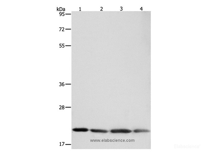
    Western Blot analysis of Human brain malignant glioma, MCF-7 cells, Raji cells and LoVo cells using BAX Polyclonal Antibody at dilution of 1:300.

    Western Blot analysis of Human brain malignant glioma, MCF-7 cells, Raji cells and LoVo cells using BAX Polyclonal Antibody at dilution of 1:300.

    Western Blot analysis of Human brain malignant glioma, MCF-7 cells, Raji cells and LoVo cells using BAX Polyclonal Antibody at dilution of 1:300.

    Western Blot analysis of Human brain malignant glioma, MCF-7 cells, Raji cells and LoVo cells using BAX Polyclonal Antibody at dilution of 1:300.

    IHC
    ( esophagus cancer,lung cancer, )

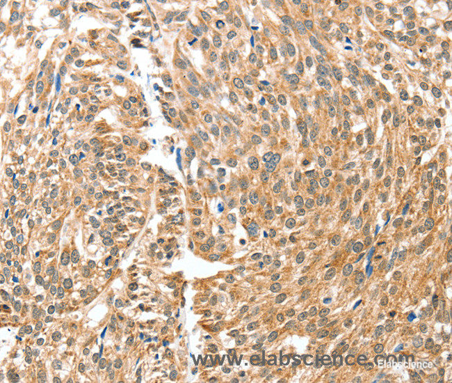
    Immunohistochemistry of paraffin-embedded Human esophagus cancer using BAX Polyclonal Antibody at dilution of 1:30.

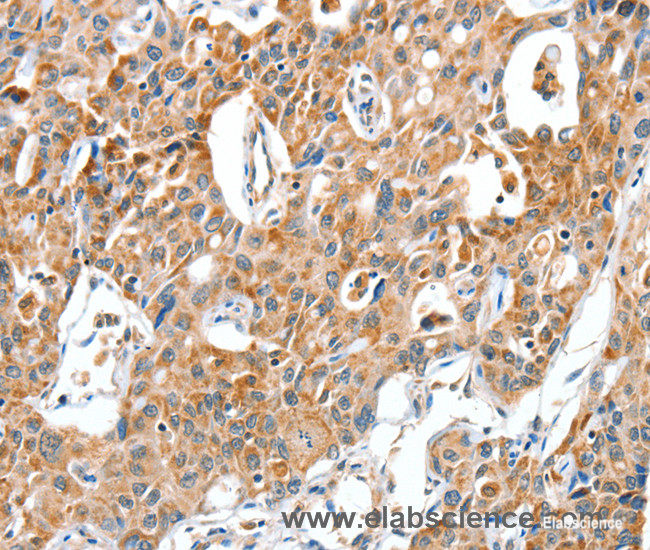
    Immunohistochemistry of paraffin-embedded Human lung cancer using BAX Polyclonal Antibody at dilution of 1:30.

  • Dilution

    WB 1:500-1:2000
    IHC 1:25-1:100

Preparation of protein samples

1.Protein extraction

1)For tissue sample
a. Take the samples, wash the tissue thoroughly with pre-cooled PBS (0.01 M, pH=7.4)(Cat# E-BC-R187) to remove the surface blood and internal debris.
b. Weigh and smash the tissue, add an appropriate ratio of RIPA Lysis Buffer (Cat# E-BC-R327)(add 10 μL PMSF and 10 μL Na3VO4 to each 1 mL RIPA Lysis) and homogenizely lyse the tissue.
It is recommended to homogenize according to the ratio of tissue weight: RIPA volume = 3:10. For example, add 1 mL RIPA Lysis Buffer to 0.3 g tissue sample, the specific volume can be adjusted according to experimental requirements.
c. Shake and lyse on the ice for 30 min after homogenization. And then sonicate the sample for 1 min (under ice water bath conditions) with 2 s’ sonication and 2 s’ intervals to make cells fully lysis and reduce the viscosity of sample.
d. Centrifuge at 12,000 rpm for 10 min at 4℃.
e. Take the supernatant and measure the protein concentration mentioned in step2.

2)For cell sample
a. Collect the cells, wash them thoroughly with pre-cooled PBS (0.01 M, pH=7.4) to remove the medium off (it is generally recommended to wash 3 times).
b. Add an appropriate ratio of RIPA Lysate Buffer (10 μL PMSF and 10 μL Na3VO4 in each 1 mL RIPA Lysis) and lyse on the ice for 30 min.
It is recommended to add 0.1 mL of RIPA Lysis Buffer to each well of a 6-well plates (the protein content in different cells may vary, and the volume of the lysate added can be appropriately adjusted).
c. Sonicate the sample for 1 min (under ice water bath conditions) with 2 s’ sonication and 2 s’ intervals to make cells fully lyse and reduce viscosity of sample.
d. Centrifuge at 12,000 rpm for 10 min at 4℃.
e. Take the supernatant and measure the protein concentration mentioned in step2.

2.Measurement of protein concentration
By the BCA method (see the Total Protein Colorimetric Assay Kit (Cat# E-BC-K318) instructions).

3.Boiling the samples
Adjust the protein concentration with PBS Buffer. Add 5 × SDS Loading Buffer (Cat# E-BC-R288) with the ratio of the protein sample: 5 × SDS Loading Buffer = 4:1 and boil the mixture for 10 min. Centrifuge at 12,000 rpm for 2 min and collect the supernatant. The denatured protein can be employed to Western Blot experiments or stored at -20℃ or -80℃.

Note: It is recommended that the total protein loading amount of test sample is about 50 μg in each well. Try to make the loading volume of each sample close to 10 μL.

Electrophoresis

1.According to the molecular weight of the target protein, prepare 0% separation gel. Add the test sample to each well, and add 5 μL of Pre-stained Protein Marker (Cat# E-BC-R273)to a reserved well in order to verify the target molecular weight and the extent of membrane transfer. Add Electrophoresis Buffer ( Cat# E-BC-R331) and start electrophoresis.

2.Electrophoresis at 80v when the samples are in stacking gel, then convert to 120v when the blue flow into the separating gel. Electrophoresis time is about 2-3 h till bromophenol blue reaches the bottom of the gel.

Transfer Membrane

1.Choose the PVDF Membrane (Cat# E-BC-R266) with a pore size of μm according to the molecular weight of the target protein. Soak the PVDF Membrane in methanol for 1 min to activate it, and then soak the PVDF Membrane in the Transmembrane Buffer (Cat# E-BC-R333), the filter paper and fiber mat must be soaked in the Transmembrane Buffer for use too.

2.Follow manufacture instructions of Transfer System for wet, semi-dry, or dry transfer.

Incubation of antibodies

1.Soak the PVDF Membrane with TBST Buffer (Cat# E-BC-R335) containing 5% Skim Milk Powder as blocking buffer and block the membrane at room temperature for .

2.According to the recommended primary antibody dilution ratio, use the TBST Buffer containing 5% Skim Milk Powder to dilute the BAX Antibody at , soak the PVDF Membrane in the primary antibody working solution, incubate overnight at 4 ℃, and gently shake.

3.Wash the PVDF Membrane with TBST Buffer for .

4.According to the recommended secondary antibody dilution ratio, use a TBST Buffer solution containing 2% Skim Milk Powder to dilute Goat Anti-Rabbit IgG (H+L) (peroxidase/HRP conjugated) (Cat# E-AB-1003) at . Incubate at room temperature for 1 h on a shaker.

5.Wash the PVDF Membrane with TBST Buffer for .

Detection

1.Mix A and B in the Excellent Chemiluminescent Substrate Detection kit (Cat# E-BC-R347) at the ratio of 1:1 as working solution.

2.Take out the PVDF Membrane from TBST Buffer and absorb the liquid with the filter paper. Pave the PVDF Membrane on the detection machine, add ECL working solution continuously on the PVDF Membrane, discharge the bubble and detect the result.

3.Adjust the contrast and the exposure time to get the best image.

Appendix

Product Details

Clonality Polyclonal
Isotype IgG
Concentration 0.2 mg/mL
Storage Store at -20℃. Avoid freeze / thaw cycles.
Buffer PBS with 0.05% sodium azide and 50% glycerol, PH7.4
Purification Method Affinity purification
Research Areas Cancer, Cell Biology, Metabolism
Conjugation Unconjugated

Immunogen Details

Immunogen Recombinant protein of human BAX
Abbre BAX
Synonyms Apoptosis regulator BAX,BAX,Bax-protein,BAX,BAXA,Baxdelta2G9,Baxdelta2G9omega,Baxdelta2omega,Bcl-2-like protein 4,BCL2 associated X protein,BCL2 associated X protein omega,BCL2 associated X protein transcript variant delta2,Bcl2-L-4,BCL2L4,membrane isoform alpha
Swissprot Q07812
Gene Accession BC014175
Calculated MW 21kDa
Cellular Localization Cytoplasm and Mitochondrion membrane. Cytoplasm. Colocalizes with 14-3-3 proteins in the cytoplasm. Under stress conditions, undergoes a conformation change that causes release from JNK-phosphorylated 14-3-3 proteins and translocation to the mitochondrion membrane.
Tissue Specificity Expressed in a wide variety of tissues. Isoform Psi is found in glial tumors. Isoform Alpha is expressed in spleen, breast, ovary, testis, colon and brain, and at low levels in skin and lung. Isoform Sigma is expressed in spleen, breast, ovary, testis, lung, colon, brain and at low levels in skin. Isoform Alpha and isoform Sigma are expressed in pro-myelocytic leukemia, histiocytic lymphoma, Burkitt's lymphoma, T-cell lymphoma, lymphoblastic leukemia, breast adenocarcinoma, ovary adenocarcinoma, prostate carcinoma, prostate adenocarcinoma, lung carcinoma, epidermoid carcinoma, small cell lung carcinoma and colon adenocarcinoma cell lines.

Background

The protein encoded by this gene belongs to the BCL2 protein family. BCL2 family members form hetero- or homodimers and act as anti- or pro-apoptotic regulators that are involved in a wide variety of cellular activities. This protein forms a heterodimer with BCL2, and functions as an apoptotic activator. This protein is reported to interact with, and increase the opening of, the mitochondrial voltage-dependent anion channel (VDAC), which leads to the loss in membrane potential and the release of cytochrome c. The expression of this gene is regulated by the tumor suppressor P53 and has been shown to be involved in P53-mediated apoptosis. Multiple alternatively spliced transcript variants, which encode different isoforms, have been reported for this gene.

Citations

  1. Biomolecules (2022) IF: 6.064
    First Evidence of the Expression and Localization of Prothymosin α in Human Testis and Its Involvement in Testicular Cancers

    DOI: 10.3390/biom12091210

    Sample: Testicular Tissue
  2. International Journal of Reproductive Biomedicine (2021)
    Follicular development and the expression of BAX and vascular endothelial growth factor in transplanted ovaries in uni- and bilateral ovariectomized mice: An experimental study

    DOI: 10.18502/ijrm.v19i4.9062

    Sample: Ovaries
  3. INTERNATIONAL MICROBIOLOGY (2018) IF: 0.796
    attenuates hippocampal apoptosis induced by lipopolysaccharide in rats

    DOI: 10.1007/s10123-018-00051-3

    Sample: Tissue Homogenate
  4. JOURNAL OF FLUORESCENCE (2023) IF: 2.525
    The Novel 5-Fluorouracil Loaded Ruthenium-based Nanocarriers Enhanced Anticancer and Apoptotic Efficiency while Reducing Multidrug Resistance in Colorectal Cancer Cells

    DOI: 10.1007/s10895-023-03180-9

    Sample: Hct116 Cell
  5. BIOCHEMICAL AND BIOPHYSICAL RESEARCH COMMUNICATIONS (2019) IF: 2.705
    Regulated rutin co-administration reverses mitochondrial-mediated apoptosis in Plasmodium berghei-infected mice

    DOI: 10.1016/j.bbrc.2019.11.067

    PMID: 31767147

    Sample: Tissue
  6. BMC Endocrine Disorders (2022) IF: 3.263
    Low-dose spironolactone ameliorates adipose tissue inflammation and apoptosis in letrozole-induced PCOS rat model

    DOI: 10.1186/s12902-022-01143-y

    PMID: 36071485

    Sample: Adipose Tissue
  7. TOXICOLOGY (2019) IF: 3.547
    Ethanol increases manganese—Induced spatial learning and memory deficits via oxidative/nitrosative stress induced p53 dependent/independent hippocampal apoptosis

    DOI: 10.1016/j.tox.2019.03.001

    Sample: Tissue
  8. LIFE SCIENCES (2022) IF: 5.037
    Suppression of PCSK9/NF-kB-dependent pathways by acetate ameliorates cardiac inflammation in a rat model of polycystic ovarian syndrome

    DOI: 10.1016/j.lfs.2022.120560

    PMID: 35452635

    Sample: Cardiac Tissue
  9. Neurotherapeutics (2019) IF: 5.552
    Galantamine Inhibits Aβ 1–42 -Induced Neurotoxicity by Enhancing α7nAChR Expression as a Cargo Carrier for LC3 Binding and Aβ 1–42 Engulfment During Autophagic Degradation

    DOI: 10.1007/s13311-019-00803-7

    Sample: Sh-Sy5Y Cell
  10. BIOMEDICINE & PHARMACOTHERAPY (2020) IF: 6.53
    Gastroprotective effect of gallic acid against ethanol-induced gastric ulcer in rats: Involvement of the Nrf2/HO-1 signaling and anti-apoptosis role

    DOI: 10.1016/j.biopha.2020.110075

    PMID: 32179202

    Sample: Tissue
Show AllShorten

Reviews/Q&A

  • Show all
  • Reviews
  • Q&A
... Show All Show Less

People Also Bought

Apply for 10 Test Free Trial FCM Antibody
*Product Name:
*Catalog Number:
*Name:
*Email:
*Message:
*Country:
*When will you use it?

*Captcha: