| Product Name |
Sizes |
Price |
| AF/LE Purified Anti-Mouse Cd3e Antibody[17A2] |
50μg, 500μg, 1mg |
Check More |
| Purified Anti-Mouse CD3 Antibody[17A2] |
25μg, 100μg |
Check More |
| Biotin Anti-Mouse CD3 Antibody[17A2] |
25μg, 100μg |
Check More |
| FITC Anti-Mouse CD3 Antibody[17A2] |
50Tests, 100Tests, 100Tests×2 |
Check More |
| PE Anti-Mouse CD3 Antibody[17A2] |
50Tests, 100Tests, 100Tests×2 |
Check More |
| APC Anti-Mouse CD3 Antibody[17A2] |
50Tests, 100Tests, 100Tests×2 |
Check More |
| PerCP Anti-Mouse CD3 Antibody[17A2] |
50Tests, 100Tests, 100Tests×2 |
Check More |
| PE/Cyanine5 Anti-Mouse CD3 Antibody[17A2] |
50Tests, 100Tests, 100Tests×2 |
Check More |
| PE/Cyanine7 Anti-Mouse CD3 Antibody[17A2] |
50Tests, 100Tests, 100Tests×2 |
Check More |
| PE/Cyanine5.5 Anti-Mouse CD3 Antibody[17A2] |
50Tests, 100Tests, 100Tests×2 |
Check More |
| PerCP/Cyanine5.5 Anti-Mouse CD3 Antibody[17A2] |
50Tests, 100Tests, 100Tests×2 |
Check More |
| Elab Fluor® 488 Anti-Mouse CD3 Antibody[17A2] |
50Tests, 100Tests, 100Tests×2 |
Check More |
| Elab Fluor® 647 Anti-Mouse CD3 Antibody[17A2] |
50Tests, 100Tests, 100Tests×2 |
Check More |
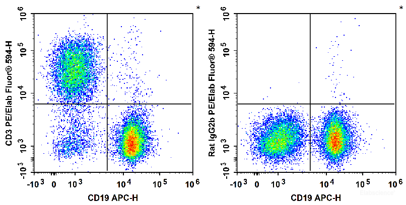
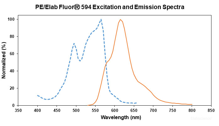
| PE/Elab Fluor® 594 Anti-Mouse CD3 Antibody[17A2] |
50Tests, 100Tests, 100Tests×2 |
Check More |
| Elab Fluor® Violet 450 Anti-Mouse CD3 Antibody[17A2] |
50Tests, 100Tests, 100Tests×2 |
Check More |
| Elab Fluor® Red 780 Anti-Mouse CD3 Antibody[17A2] |
50Tests, 100Tests, 100Tests×2 |
Check More |
| FITC Anti-Mouse CD3 Antibody[17A2] |
25μg, 100μg |
Check More |
| PE Anti-Mouse CD3 Antibody[17A2] |
25μg, 100μg |
Check More |
| APC Anti-Mouse CD3 Antibody[17A2] |
25μg, 100μg |
Check More |
| PerCP Anti-Mouse CD3 Antibody[17A2] |
25μg, 100μg |
Check More |
| PE/Cyanine5 Anti-Mouse CD3 Antibody[17A2] |
25μg, 100μg |
Check More |
| PE/Cyanine7 Anti-Mouse CD3 Antibody[17A2] |
25μg, 100μg |
Check More |
| PE/Cyanine5.5 Anti-Mouse CD3 Antibody[17A2] |
25μg, 100μg |
Check More |
| PerCP/Cyanine5.5 Anti-Mouse CD3 Antibody[17A2] |
25μg, 100μg |
Check More |
| Elab Fluor® 488 Anti-Mouse CD3 Antibody[17A2] |
25μg, 100μg |
Check More |
| Elab Fluor® 647 Anti-Mouse CD3 Antibody[17A2] |
25μg, 100μg |
Check More |
| Elab Fluor® Violet 450 Anti-Mouse CD3 Antibody[17A2] |
25μg, 100μg |
Check More |
| Elab Fluor® Red 780 Anti-Mouse CD3 Antibody[17A2] |
25μg, 100μg |
Check More |
